|
Objective
Independent:- Surface area:Volume ratio Dependent:- Rate of diffusion 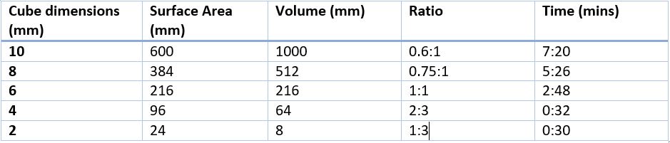 Questions:
1. What predictions did you make about the rate of diffusion and the effect of surface area to volume ration? I predicted that as the surface area to volume ratio increased so would the rate of diffusion as less time would be taken for the solution to diffuse through the agar cube. 2. Identify at least three key features you controlled in this experiment. The temperature, concentration of acid and volume of acid were three of the controlled variables that we kept constant for our experiment. 3. What additional procedures could you carry out to make these results more reliable and accurate? Repeating the experiment and taking averages would help increase the reliability. To increase the accuracy of the experiment a machine could have been used to more accurately cut the cubes reducing the human error in cutting them and increasing accuracy. 4. Explain the effect of surface area to volume ration on the rate of diffusion and how this is important in living organisms. Surface area to volume ratio is important in small organisms as it allows them to diffuse gases directly into their respiring cells. For organisms that do not have a respiratory system they use diffusion to get gases to their cells. This is because they have a large surface area and a small volume inside for the gases to reach. So the reduced diffusion distance and low metabolic activity means that they can get enough gases to the cells using diffusion.
0 Comments
This was an experiment designed to demonstrate the effects of osmosis on a cell, in this case an onion cell. Method:
The onion cells soaked in water The onion cells soaked in sucrose We also used this experiment as an opportunity to practising our microscopy work by measuring the cells using a graticule and a rulered eye piece. 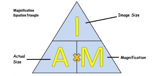 We can use this equation to work out the actual size of our cell. We can calculate the image size from the graticule in the lens. and then once we know this we can measure the cell using the ruler on the eyepiece and use the equation to work out the actual size of the cell.
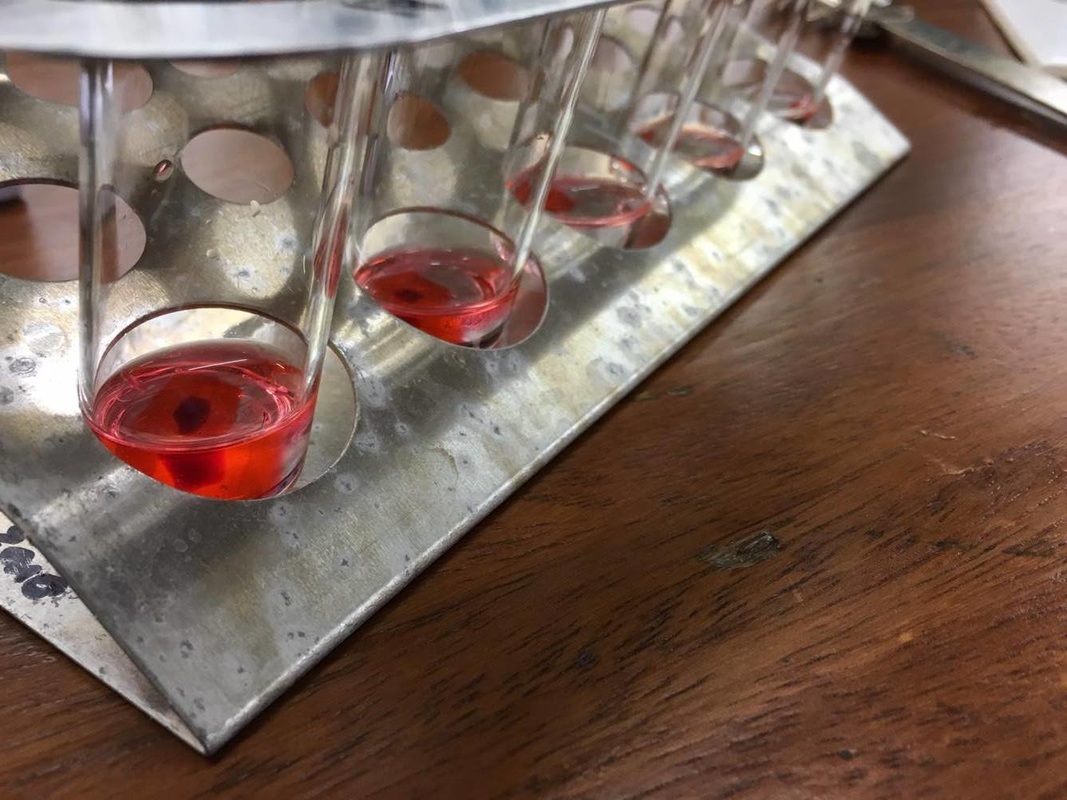
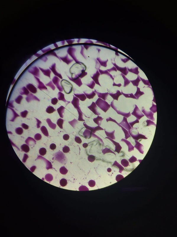
Blood staining, why we do it: Equipment:
Method:
We stain blood samples so as to stain the nucleus of the various cells we are interesting in looking at in the blood, since we are staining the nucleus and red blood cells do not contain a nucleus this clearly shows the white blood cells we are interested in. Argyroneta aquatica, a species of underwater spider which breathe through an air bubble that they trap underwater using fine hairs on their abdomens and was first described by scientist of 250 years ago has finally had the secret to its ability to remain underwater for over 24 hours without replenishing their air bubble revealed, by scientist Roger Seymour from the University of Adelaide in South Australia and Stefan Hetz of Humboldt University in Berlin, Germany. Using an experiment where they placed 12 spiders in individual aquaria and measured the oxygen levels within the air bubbles using optical fibres tipped with oxygen-sensitive dye, they discovered that the spiders web actually acted as a form of gills and diffused oxygen from the surrounding water as well as removing carbon dioxide from the bubble allowing them to stay underwater for such extraordinary lengths of time, it isn't until the nitrogen build up in the bubble is too much that they have to go to the surface and renew their bubble. Cor Vink, an entomologist at biosecurity firm AgResearch, based in Christchurch, New Zealand. sums it up by saying “It shows how amazing and versatile spider webs can be.” Review of an article from the 'New Scientist' https://www.newscientist.com/article/dn20557-scuba-spider-uses-web-as-gill-to-breathe-underwater/ 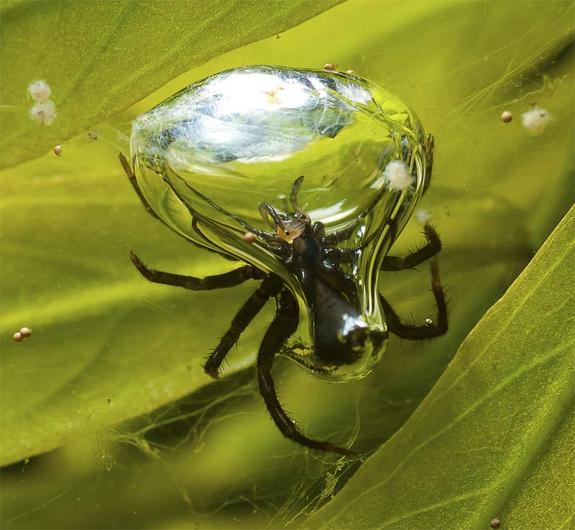 Humans have a double circulatory system meaning that there is a system called the pulmonary circuit which takes deoxygenated blood from the heart to the lungs and then takes the oxygenated heart back from the lungs to the heart. As well as this there is the systemic circuit which takes oxygenated blood from the heart all around the body to the respiring cells and then takes the deoxygenated blood back to the heart.  Equipment: Scalpel Scissors Tweezers One heart Syringe Ink Gloves Cutting board Aim: We dissected a heart to study its internal structures such as the ventricles, atria and semilunar valves. |
Jack BojanArdingly College Archives
March 2017
|


 RSS Feed
