|
Health and safety assessment table: 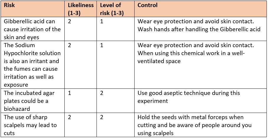 Equipment:
Background: A cereal grain contains a store of starch within the endosperm. During germination the starch must be made soluble so that it can be transported to the embryo to support the growth of the seedling. The embryo is much smaller than the endosperm and is situated at the more pointed end of the grain. The developing embryo releases gibberellins that act on a layer of cells on the outside of the endosperm, stimulating these cells to release the starch-digesting enzyme amylase. "Gibberellic acid does not merely trigger α-amylase synthesis, but it is continuously required during the period of enzyme formation" (1). In this activity you will remove the embryo and investigate the effect of different concentrations of gibberellin on the production of amylase. The production of amylase will be assessed by using a starch agar assay. Cereal grains that have had the embryo removed are first soaked in gibberellic acid, then placed onto the starch agar plates and incubated. The agar plate is then flooded with iodine solution, which stains starch blue-black. The areas where starch has been digested will not stain. The size of the clear area around a cereal grain indicates the amount of amylase produced by the seed. Variables: Independent variable:- Concentration of the Gibberellic acid Dependent variable:- Size of the zone of exclusion. This will be attained placing the zone over a sheet split up into 1mm by 1mm squares and counting the squares that fit into the zone to work out the area in mm^2 Controlled variables:-
1 Comment
The GFP gene which codes for a green fluorescent protein is isolated from the species of jellyfish Aequorea Victoria. This GFP is combined with 2 other genes; The Bla gene which codes for resistance to beta-lactam antibiotics such as ampicillin and the gene araC which is a promoter region that regulates the expression of GFP meaning that the GFP gene will only be expressed in the presence of arabinose. This DNA Method:
Results:
Health and safety assessment table 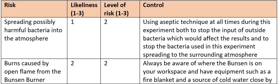 Variables: Independent variable:- Concentration of caffeine given to each Daphnia (0, 0.001, 0.01, 0.1, 1, 10, 100) ml/L Dependent variable:- The heartrate of the Daphnia measured in beats per minute Controlled variables:-
Apparatus:
Method:
Risk assessment: Use of glassware - Glassware may shatter when being used and create glass shards that can cause damage to you or others. Risk level 2 as glassware has the potential to cause serious harm. Risk likeliness 1, if used correctly and sensibly the glassware should remain intact and even if it does shatter it will most likely not injure anyone. Results: Equipment:
Method:
 Diagram credits go to Toby Brann  A good example of pollen tube growth. 0.2 sucrose solution at 15:30 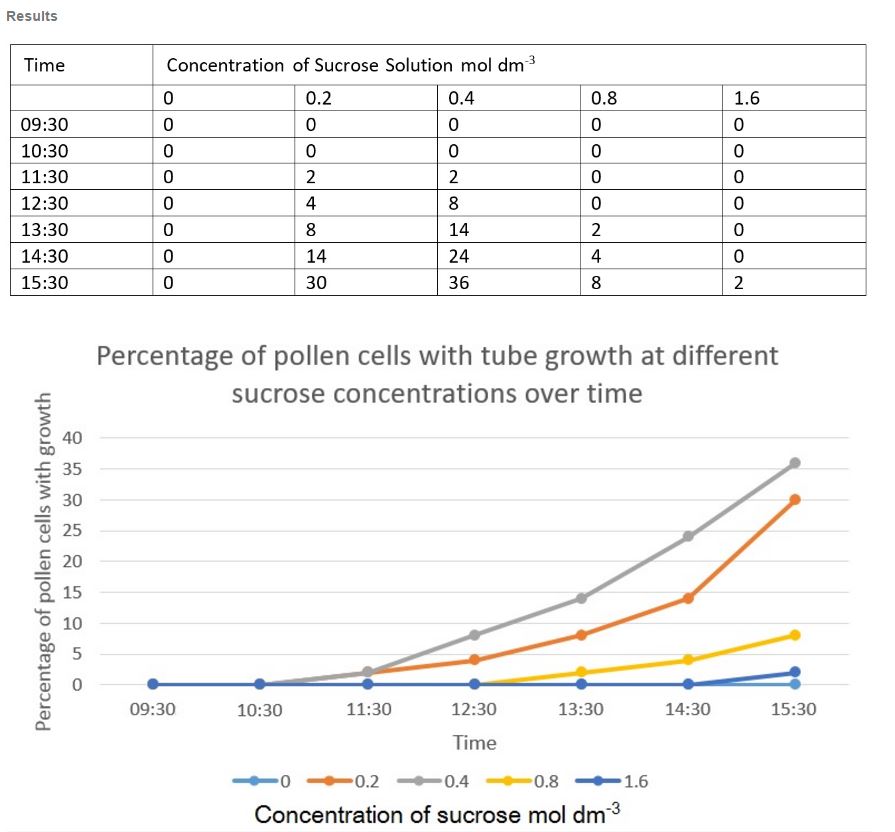 Analysis:
The graph shows us that the most effective concentration for pollen tube growth was 0.4 mol dm^3 as the percentage of pollen cells with a tube formed was 36% compared to 30% of 0.2 mol dm^3. This expreiment took place over 6 hours (09:30 - 15:30) and as we can see from our results the pollen grains in the solution containing no sucrose experienced no growth at all throughout the 6 hours period. However, the solutions with 0.8 and 1.6 mol dm^3 sucrose concertration showed relatively little growth with 8% and 2% respectively. The only difference between all the tests was the sucrose concentration that the pollen was kept in for during the experiment. Therefore differences in observations would be due to this difference in sucrose concentration. We can see that the for the higher sucrose concentrations of 1.6 and 0.8 the pollen tubes have severely stunted growth due to the fact that water travels from pollen to the surrounding through osmosis. This is due to the area surrounding the pollen being a low concentration of water. For pollen in a solution of no sucrose the pollen is in a low water concentration so, by osmosis, the water travels from the surroundings into the cytoplasm of the cell. This may end up lysing the cell if the concentration difference is significant enough. It appears, from our experiment that the concentration within the cell and the surroundings are similar enough, in the cases of 0.2 and 0.4 mol dm^3 sucrose solutions, that the pollen tubes can grow and form. 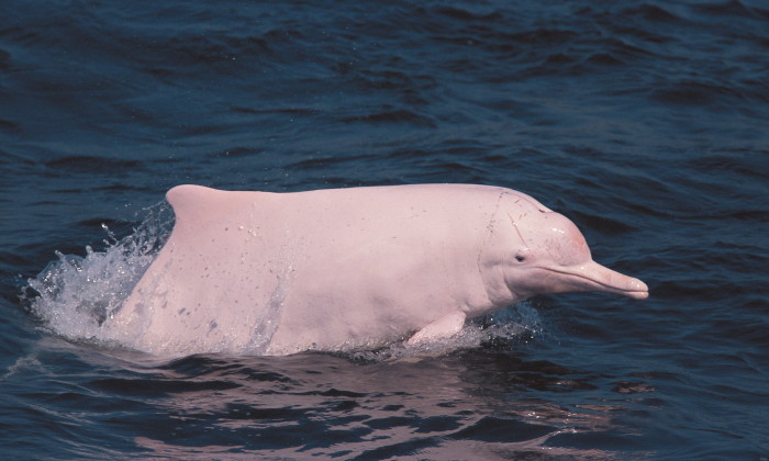 The 'Pink Dolphin' Is actually a species of dolphin known as the Chinese white dolphin, adult dolphins of this species living along the Chinese coast however have developed pink skin. This is thought to be caused by blood vessels that were overdeveloped for thermoregulation. The 'Pink Dolphin' Is a species very close to my heart as as its existence is tied in with Hong Kong and it has become a symbol of the city and was first seen as a bad omen in the times when Hong Kong was a small fishing community and it was thought that these dolphins would steal fish from the nets, they are now however seen as a natural treasure that we are unfortunately losing due to land reclamation, pollution and the construction of structures such as a 50km bridge linking Hong Kong to Macau. The estimated population of pink dolphins living in Hong Kong waters was recorded as 61 dolphins as of December 2015. Kingdom: Animalia Phylum: Chordata Class: Mammalia Order: Artiodactyla Family: Delphinidae Genus: Sousa Species: Chinensis  The blue in this map highlights the areas in which this Dolphin is thought to be found. However as mentioned previously they are only pink along the coastal waters of China and Hong Kong. Conservation of the Pink Dolphin In 2003 There was an estimated 158 Pink Dolphins left in the waters of Hong Kong, and now there is an estimated 61 Dolphins remaining. That is a 60% decrease in just over a decade. In 2010 alone 17 dead dolphins were found washed up along the beaches of Hong Kong and although of course death is natural, these Dolphins have a lifespan of 40 years and only reproduced once a year, so a death of 17 is highly significant. There are more than a couple of reasons why this species is dying out, all of which are caused by humans. One is to do with pollution. Sewage ends up in the water, and the dolphins swim through it. As they swim through this polluted water, it enters their bodies. When female dolphins feed their young with their milk, the milk contains absorbed toxins from the water. This process is explained further below. This process of toxins being built up within organisms is a process called bioaccumulation. This is when an animal ingests a contaminated item and the toxin stays in its body. However the dolphins most likely ingest the toxins through biomagnification which is when a predator eats prey infected with a toxin and becomes infected itself. In the ocean, when pollutants like polychlorinated biphenyls (PCBs), enter the water, the first organisms affected are often simpler ones like plankton or algae. The toxins enter a dolphin’s body when it ingests contaminated fish or simpler organisms. Researchers have discovered that instead of eliminating the toxins in its waste, a dolphin stores the offending chemical in its fatty tissues, or blubber, and breast milk. These chemicals in the breast milk can then go on to cause harmful and even possibly fatal birth defects to the calfs which directly affects the pink dolphin population. As well as sewage in the water, a large portion of Hong Kong's rubbish is thrown into the sea every year and this floating debris can trap, maim or even drown the dolphins by not allowing them to reach the surface to acquire oxygen. Another problem is overfishing. Dolphins only eat fish. This is a huge problem in Hong Kong because we take too many fish out of the sea, meaning there are hardly any left for the dolphins to eat. A huge effort will have to put in by the Hong Kong people and also internationally if this species that is classified on the IUCN red list as critically endangered is to fight off extinction. Luckily already in Hong there is a commpany www.hkdolphinwatch.com/ who have regular boat excursions to observe the Dolphins which helps raise awereness for this endagered species and hopefully encorauage people to start preserving our waters and the wildlife within them. "It is up to the government and every Hong Kong citizen to stand up for dolphins. We risk losing them unless we all take action," - Samuel Hung, Society Chairman 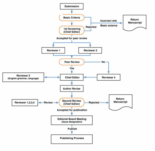 A flowchart to demonstrate how the peer review process works. Below is a link to my research paper written by me and fellow student Adam Housby
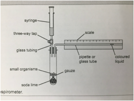
 CPAC 9: Investigating factors affecting the rate of aerobic respiration using a respirometer12/10/2016 Apparatus:
Safety: Soda lime is highly corrosive so eye protection should be worn at all times and contact with the soda lime should be avoided. A spatula should be used to handle it. After handling the maggots you should wash your hand thoroughly. Procedure:
Results: 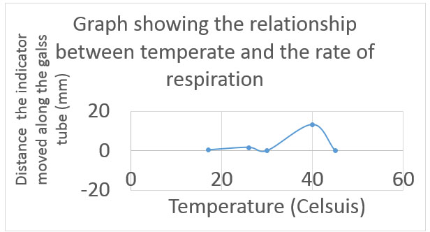 As we can see from these results, overall as the temperature increased, so did the rate of respiration measured by the distance the indicator moved along the glass tube in a minute (measured in mm). However the optimum temperature seemed to be 40 degrees Celsius and after the point the rate of respiration began to decrease again until at 50 degrees Celsius it was at 0.
The rate of respiration actually reached 0 at 55 degrees Celsius because the temperature turned out to be too hot for the maggots and they perished under the conditions which was very unethical and for future experiments lower temperatures needs to be used to ensure the protection of the organisms. Reticulated pythons or python reticulatus are a species of snake found in Southern Asia and are part of the python genus meaning they are nonvenomous and kill there prey by constriction and asphyxiation. They are considered as the world's longest (although to the bulkiest) snake with the largest accurately recorded length to be of a snake known as Medusa who measured in at an incredible 7.67 m (25 ft 2 in) long [1] although there have been reports of snakes reaching 10.1 m (33 ft 1 in). These snakes have been suggested to be one of the few snakes who can actually target humans as prey and have been linked to a number of human fatalities. 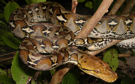 The Respiratory system Snakes have a small opening just behind the tongue called the glottis, which opens into the trachea. Unlike what mammals have, the reptile glottis is always closed, forming a vertical slit, unless the snake takes a breath. A small piece of cartilage just inside the glottis vibrates when the snake forcefully expels air from its lungs. This produces a snake’s characteristic hiss. Snakes are able to extend their glottis out the side of their mouth while they eat, which allows for respiration while they consume large prey items. 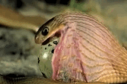 The trachea is a long, straw-like structure supported by cartilaginous rings. These rings are incomplete in that the snake looks more like a C than an O. A thin membrane completes the open part of the C. This configuration is also seen in lizards, but the function of the incomplete rings remains unknown. The trachea usually terminates just in front of the heart, and at this point it splits into the two primary bronchi, airways that direct air into either the left or right lung. In most snakes the short left bronchus terminates into a rudimentary left lung and only the right lung is used. however in the case of Boas and Pythons such as our Reticulated Python both lungs are used. The left lung can also be used by water snakes but as an aid for buoyancy rather than for respiration. 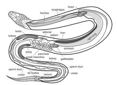 Snakes breathe principally by contracting muscles between their ribs. Unlike mammals, they lack a diaphragm. Inspiration is an active process (muscles contract), whereas expiration is passive (muscles relax). The portion of a snake’s lung nearest its head has a respiratory function; this is where oxygen exchange occurs. The lung portion nearest the tail, regardless of the lung’s size, is more of an air sac. The inside of these sac portions look more like the inside of a balloon than a lung. There is no exchange of respiratory gases.
Procedure:
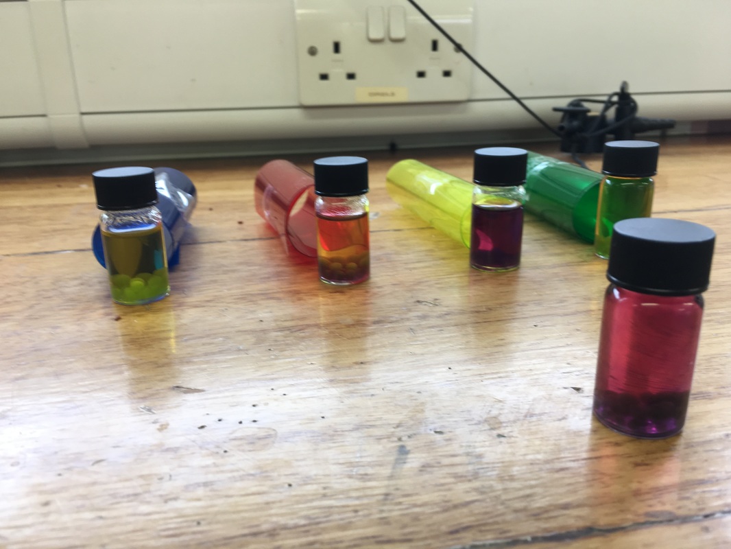
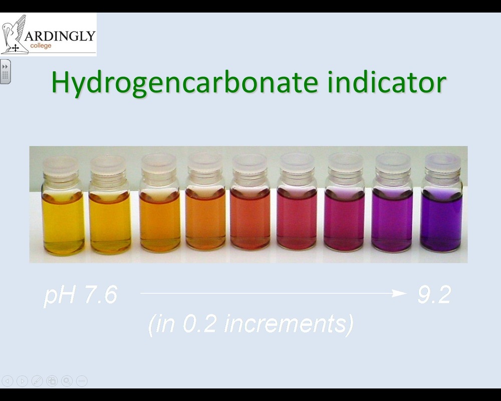
Results: The image on the top left is the results I obtained from carrying out this experiment and the photo on the top right is a scale showing how the hydrogen carbonate indicator reacts to different pH levels. As we can see the glass vial that was left out without a colored filter around it turned a deep purple color and from the scale this suggest that the pH in that vial was around 8.5. This is because since all wavelengths of light were allowed into the vial so that the alga in their could photosynthesize to their full ability and the production of CO2 coming from that photosynthesis is what caused the change of pH within the vial. However as we can see with the vial which had the green filter on, the color has stayed yellow suggesting a pH of around 7.9. This is because that the green filter was inhibited all wavelengths of light other than those in the green spectrum. As we can see on the below graph, the pigments within the chloroplasts responsible for photosynthesis are unable to absorb light in the green spectrum (around 520-550 nm) and since the alga cannot photosynthesis, CO2 isn't produced to alter the pH level of the solution like with the vial without the filter. 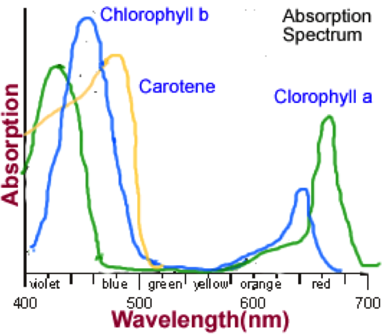 |
Jack BojanArdingly College Archives
March 2017
|
||
 RSS Feed
