|
Aim: Investigation into the effect of temperature on membrane permeability Apparatus:
Method:
Health and Safety:
The below three photos are credited to Henry Gould 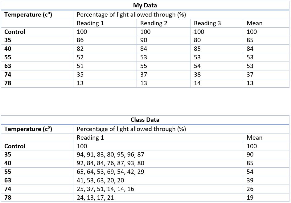  Analysis of results:
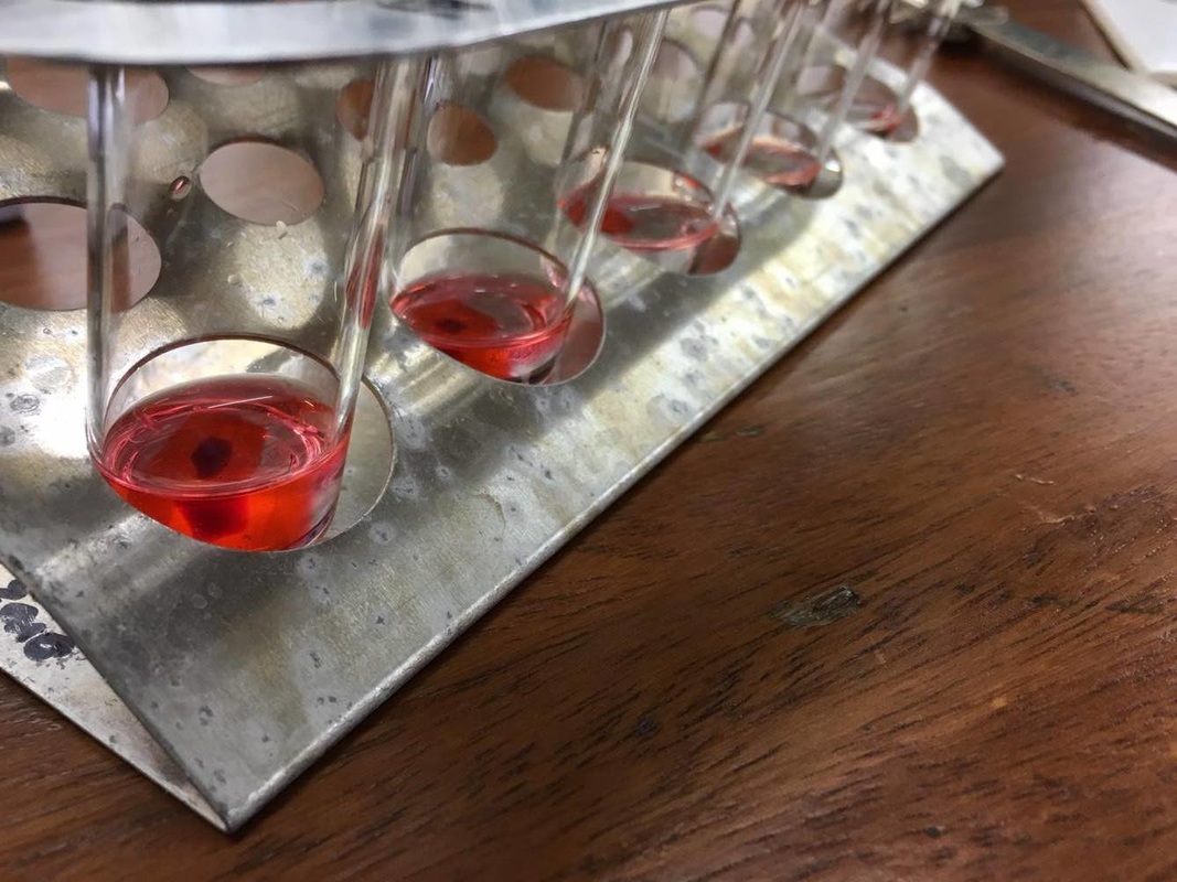
My class and I found that our graphs showed a negative correlation. This means that as the temperature increased, the transmission or percentage of light allowed through the solution decreased (visually this means the solution got darker). This is because more pigment has been leaked from the beetroot into the surrounding solution. This is because the higher temperatures have caused the plasma membrane and tonoplast of the beetroot cells to move around more from the increase in kinetic energy and for some vital membrane proteins to denature allowing more pigment out of the vacuole and then out of the cell. Questions:
5 Comments
Objective
Independent:- Surface area:Volume ratio Dependent:- Rate of diffusion 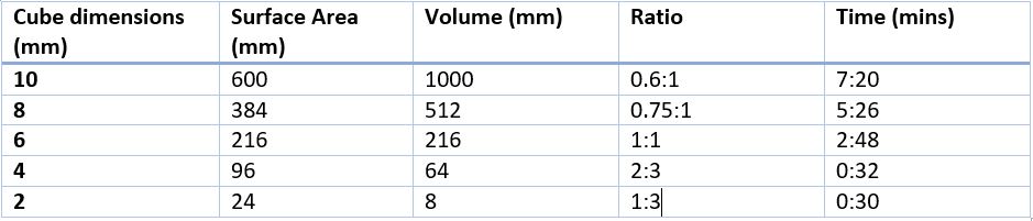 Questions:
1. What predictions did you make about the rate of diffusion and the effect of surface area to volume ration? I predicted that as the surface area to volume ratio increased so would the rate of diffusion as less time would be taken for the solution to diffuse through the agar cube. 2. Identify at least three key features you controlled in this experiment. The temperature, concentration of acid and volume of acid were three of the controlled variables that we kept constant for our experiment. 3. What additional procedures could you carry out to make these results more reliable and accurate? Repeating the experiment and taking averages would help increase the reliability. To increase the accuracy of the experiment a machine could have been used to more accurately cut the cubes reducing the human error in cutting them and increasing accuracy. 4. Explain the effect of surface area to volume ration on the rate of diffusion and how this is important in living organisms. Surface area to volume ratio is important in small organisms as it allows them to diffuse gases directly into their respiring cells. For organisms that do not have a respiratory system they use diffusion to get gases to their cells. This is because they have a large surface area and a small volume inside for the gases to reach. So the reduced diffusion distance and low metabolic activity means that they can get enough gases to the cells using diffusion. This was an experiment designed to demonstrate the effects of osmosis on a cell, in this case an onion cell. Method:
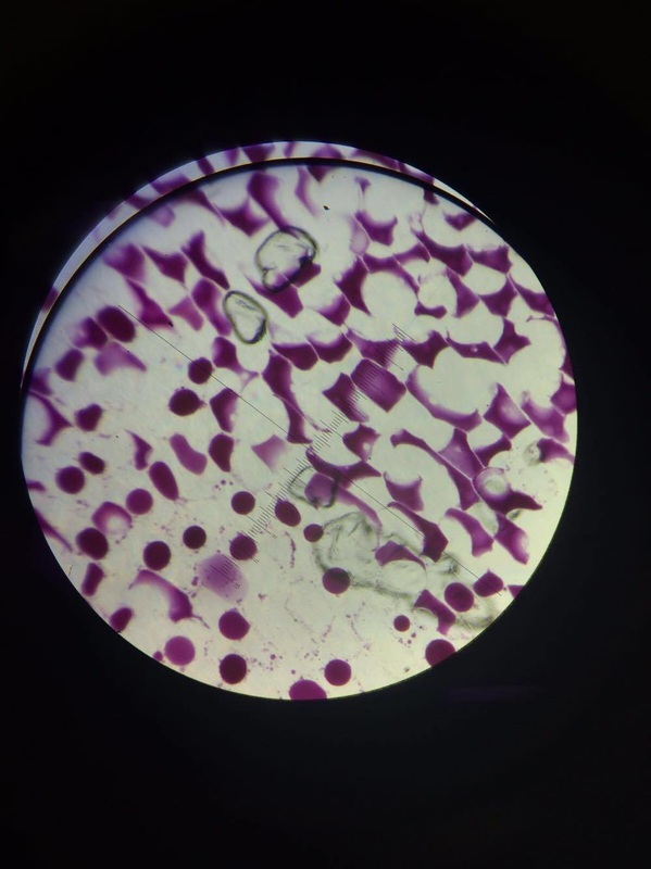
The onion cells soaked in water The onion cells soaked in sucrose We also used this experiment as an opportunity to practising our microscopy work by measuring the cells using a graticule and a rulered eye piece. 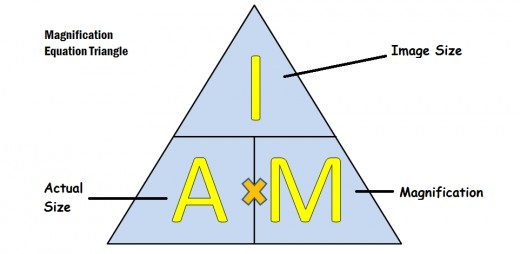 We can use this equation to work out the actual size of our cell. We can calculate the image size from the graticule in the lens. and then once we know this we can measure the cell using the ruler on the eyepiece and use the equation to work out the actual size of the cell.
Blood staining, why we do it: Equipment:
Method:
We stain blood samples so as to stain the nucleus of the various cells we are interesting in looking at in the blood, since we are staining the nucleus and red blood cells do not contain a nucleus this clearly shows the white blood cells we are interested in. Argyroneta aquatica, a species of underwater spider which breathe through an air bubble that they trap underwater using fine hairs on their abdomens and was first described by scientist of 250 years ago has finally had the secret to its ability to remain underwater for over 24 hours without replenishing their air bubble revealed, by scientist Roger Seymour from the University of Adelaide in South Australia and Stefan Hetz of Humboldt University in Berlin, Germany. Using an experiment where they placed 12 spiders in individual aquaria and measured the oxygen levels within the air bubbles using optical fibres tipped with oxygen-sensitive dye, they discovered that the spiders web actually acted as a form of gills and diffused oxygen from the surrounding water as well as removing carbon dioxide from the bubble allowing them to stay underwater for such extraordinary lengths of time, it isn't until the nitrogen build up in the bubble is too much that they have to go to the surface and renew their bubble. Cor Vink, an entomologist at biosecurity firm AgResearch, based in Christchurch, New Zealand. sums it up by saying “It shows how amazing and versatile spider webs can be.” Review of an article from the 'New Scientist' https://www.newscientist.com/article/dn20557-scuba-spider-uses-web-as-gill-to-breathe-underwater/ 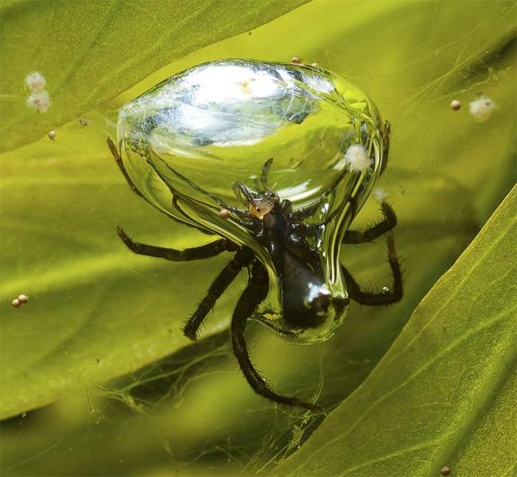 Humans have a double circulatory system meaning that there is a system called the pulmonary circuit which takes deoxygenated blood from the heart to the lungs and then takes the oxygenated heart back from the lungs to the heart. As well as this there is the systemic circuit which takes oxygenated blood from the heart all around the body to the respiring cells and then takes the deoxygenated blood back to the heart.  Equipment: Scalpel Scissors Tweezers One heart Syringe Ink Gloves Cutting board Aim: We dissected a heart to study its internal structures such as the ventricles, atria and semilunar valves. 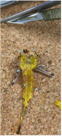 Aim Our aim was to dissect a locut to observe the inner workings and gain a better understanding of the respiratory system of locust and insects by examining the;
Equipment
Safety Concerns Tale caution when handling sharp tools such as the scalpel, Scissors and Pins and make sure not to cut yourself or anyone else. Also you may want to wear gloves or wash your hands regularly to reduce the risk of contracting any disease the dead insect may have. Method of dissection
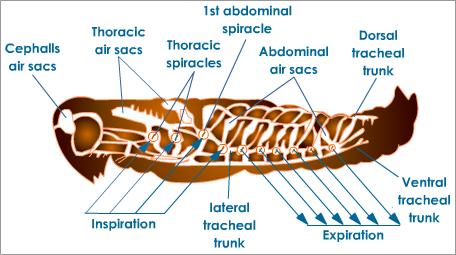
Insect Respiratory System
Air enters the insect's body through valve-like openings in the exoskeleton. These openings (called spiracles) are located laterally along the thorax and abdomen of most insects. Air flow is regulated by small muscles that operate one or two flap-like valves within each spiracle, contracting to close the spiracle, or relaxing to open it. After passing through a spiracle, air enters a tracheal, eventually diffusing throughout a complex, branching network of tracheal tubes that divide into smaller and smaller diameters and reach every part of the body. At the end of each tracheal branch, a special cell (the tracheae) provides a thin, moist interface for the exchange of gasses between atmospheric air and a living cell. Oxygen in the tracheal tube first dissolves in the liquid of the tracheole and then diffuses into the cytoplasm of an adjacent cell. At the same time, carbon dioxide, produced as a waste product of cellular respiration, diffuses out of the cell and, eventually, out of the body through the tracheal system and out through the spiracles.  Our Model We created a popular representation of the human breathing system from a plastic bottle, two balloons. a straw and some modelling clay 2. A smaller diameter straw would restrict how much air could go into out 'lung'. this is related to people with asthma to due to fact that when they have an asthmatic attack there trachea contracts and this give it a smaller diameter to the trachea restricting the airflow and therefore hampering their ability to breath. people suffering from an asthmatic attack therefor to to take medicine such as ventolin to help expand their trachea. 3. To improve this model I would use two balloons as the internal lungs to give a much more accurate visualization of the lungs and how much air is taken in by them. I would also try and use a more malleable material to create the ribcage out of so that we might be able to see the 'ribcage' move in and out with the lungs. 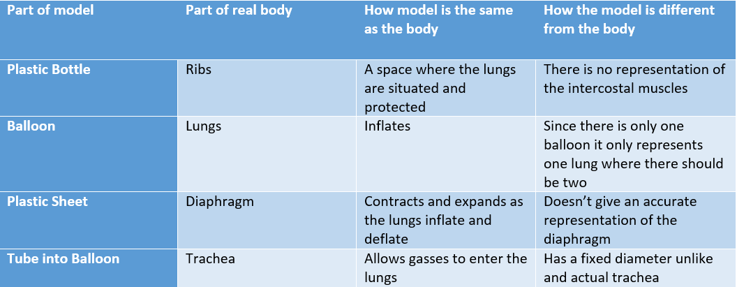 Method: 1. Prepare a broth inoculated with yeast bacteria. 2. Using a control of 1 part uninoculated broth mixed with 9 parts water, set the colorimetre. 3. Prepare a solution of 1 part yeast broth (make sure you shake the broth) and 9 parts water. Test this solution in the calorimeter for absorbency 4. Repeat each individual test three times and take an average which you will record 5. Repeat step 2-4 three times a day (morning, afternoon, evening) for 3 days More light should be absorbed as your bacteria reproduce giving you a higher level of absorbency. Results: The two results which are missing were anomalous results caused by our research group not thoroughly shaking the yeast solution before testing it which meant that a high concentration of the yeast was at the bottom and not distributed evenly affecting the results.
To isolate cholera from for example a fecal specimen, it is first enriched in an enrichment broth known as Alkaline peptone water or APW. it is grown in the APW for around 6 to 8 hours. It is then grown in the medium of choice which is usually Thiosulfate citrate bile salts sucrose agar or TCBS. TCBS is commonly used to isolate vibrio cholera as it specializes in isolating vibrio bacteria and is also very easy to produce and commercially available. |
Jack BojanArdingly College Archives
March 2017
|


 RSS Feed
